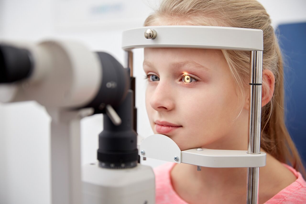
How the Eye Works
Our ability to “see” starts when light reflects off an object at which we are looking and enters the eye. As it enters the eye, the light is unfocused. The first step in seeing is to focus the light rays onto the retina, which is the light sensitive layer found inside the eye. Once the light is focused, it stimulates cells to send millions of electrochemical impulses along the optic nerve to the brain. The portion of the brain at the back of the head interprets the impulses, enabling us to see the object.
Light entering the eye is first bent, or refracted, by the cornea — the clear window on the outer front surface of the eyeball. The cornea provides most of the eye’s optical power or light-bending ability.
After the light passes through the cornea, it is bent again — to a more finely adjusted focus — by the crystalline lens inside the eye. The lens focuses the light on the retina. This is achieved by the ciliary muscles in the eye changing the shape of the lens, bending or flattening it to focus the light rays on the retina.
This adjustment in the lens, known as accommodation, is necessary for bringing near and far objects into focus. The process of bending light to produce a focused image on the retina is called “refraction”. Ideally, the light is “refracted,” or redirected, in such a manner that the rays are focused into a precise image on the retina.
Most vision problems occur because of an error in how our eyes refract light. In nearsightedness (myopia), the light rays form an image in front of the retina. In farsightedness (hypermetropia), the rays focus behind the retina. In astigmatism, the curvature of the cornea is irregular, causing light rays to focus to more than one place so that a single clear image cannot be formed on the retina, resulting in blurred vision. As we age, we find reading or performing close-up activities more difficult. This condition is called presbyopia, and results from the crystalline lens being less flexible, and therefore less able to bend light.
Since changing the apparent refraction of the eye is relatively easy through the use of corrective spectacle or contact lenses, many of the conditions that contribute to unclear vision can be readily corrected.
Sensory Interpretation:
Even with the light focused on the retina, the process of seeing is not complete. For one thing, the image is inverted, or upside down. Light from the various “pieces” of the object being observed stimulate nerve endings – photo receptors or cells sensitive to light – in the retina.
Rods and Cones:
Two types of receptors – rods and cones – are present. Rods are mainly found in the peripheral retina and enable us to see in dim light and to detect peripheral motion. They are primarily responsible for night vision and visual orientation. Cones are principally found in the central retina and provide detailed vision for such tasks as reading or distinguishing distant objects. They also are necessary for color detection. These photo receptors convert light to electrochemical impulses that are transmitted via the nerves to the brain.
Millions of impulses travel along the nerve fibers of the optic nerve at the back of the eye, eventually arriving at the visual cortex of the brain, located at the back of the head. Here, the electrochemical impulses are unscrambled and interpreted. The image is re-inverted so that we see the object the right way up. This “sensory” part of seeing is much more complex than the refractive part – and therefore is much more difficult to influence accurately.
Many optometrists are expanding their traditional role to include other areas that affect eye health, such as nutrition. Research has shown that nutrition can impact the development of cataracts and age-related macular degeneration (AMD), which are the two leading causes of blindness and visual impairment among millions of aging Americans. Nutrition may be particularly important given that currently, treatment options after diagnosis for these eye diseases are limited.
Vision and aging: a guide to good eye health and vision
Eyes often benefit from having more than one pair of prescription eyewear to meet special vision requirements. Your optometrist understands the special demands of aging and will offer specific recommendations so you can enjoy clear and comfortable vision.
As your golden years approach, even as early as 40 years old, it is especially important to make regular eye examinations part of your plan for maintaining good health and vision, as this can often delay or reduce effects of aging in our eyes. It’s also important to note that many medications to treat health conditions (like arthritis, diabetes and high blood pressure) can affect the eyes and vision, so regular treatment is vital.
Some changes in vision should be expected, even with the best preventative efforts. But as you age, there are a few common conditions you and your optometrist need to look for. Here is a short list of the most common and troubling conditions:
Presbyopia is very common among this age group. It is the loss of ability to change focus from far to near. It is often the first wake-up call that our eyes “aren’t what they used to be”. The most common signs or symptoms include the tendency to hold reading materials at arm’s length, blurred vision at normal reading distance and eye fatigue when attempting to do close work.
Glaucoma can result when excessive fluid pressures damage the optic nerve. It is one of the leading causes of blindness in Canada. Glaucoma can be effectively treated with prescription eye drops, and in some cases, surgery may be required. A simple and painless procedure allows your optometrist to measure the internal pressures of your eye. Early detection is the key to success when fighting glaucoma. Most glaucomas offer no pain or symptoms.
Cataracts are another common condition you may encounter. Cataracts occur as the lens becomes cloudy, distorting our vision. Cataracts are most often found in persons over the age of 55, but can occur in younger people as well. This condition often requires a corrective lens change or surgical removal. After surgery, you, along with your Optometrist, can decide on the best type of vision correction for you.
Macular Degeneration is a disease that obscures a person’s central field of vision.
It is the leading cause of vision loss and blindness for seniors in North America. Early detection is the key to managing the disease – that’s why yearly exams with your Optometrist are recommended.
Computers and Your Vision

With so many of us spending time in front of the computer every day it’s no surprise that research is showing a rise in visual problems. What can you do? First, it’s important to find out how you can protect your eyes through eye health exams and by making a few minor changes in your computer viewing habits.
Being far or near-sighted, having astigmatism can make computer use less comfortable and efficient. Depending on your condition, your eyes could exert extra focusing effort or be forced to work harder to maintain a clear image on the screen. The results are eye strain and fatigue. Special anti-glare coated lenses are designed to cut the blue-violet wavelength of light emitted by computer monitors and digital devices. This blue-violet light has been shown to increase eyestrain and fatigue.
We need to know:
Helpful tips to take the sting out of computer use
Correct positioning of your computer, keyboard and typing copy is essential. Your screen should be positioned about an arm’s length from your eyes and 20 degrees below eye level. Consider foot and wrist rests for added comfort.
Room lighting should be diffuse, not direct, to reduce glare and reflections from your screen. Look into an internal or external glare screen and be sure to set the color, contrast and brightness levels to suit you.
Anti-reflective coatings on the lenses of your glasses can be applied to reduce discomfort. And don’t forget, your doctor of optometry can talk to you about eyeglasses designed specifically for people who use computers a lot.
Taking a break from your work isn’t just a nice idea, it’s essential to the health and comfort of your eyes. Optometrists recommend the 20-20-20 rule… every 20 minutes take a 20 second break and focus your eyes on something at least 20 feet away (the coffee machine possibly!). This will give your eyes a much-needed break and reduce some of the symptoms mentioned earlier.
Did you know that on average we blink 12 times per minute? But wait, did you know that when we’re on the computer we only blink 5 times per minute? That can add up to dry eyes. Relieve the discomfort by using artificial tear drops or gels and remember to blink!
Common Eye Disorders
There are several common eye diseases and disorders that can cause impairment. It’s important to schedule regular eye examinations for prevention. Common disorders include:
Amblyopia is the lack of development of vision in one eye that is not directly caused by any eye health problem. It is not correctable with lenses alone. It is the result of poor early development, and as such, occurs before the age of six. It occurs in an estimated 2-4% of children under the age of six. It is caused by a large difference in the prescription between the two eyes or it can occur when strabismus (crossed eyes) is present. It can also occur when something is interfering with the clarity of the various components of the eye. This causes blurred vision in the affected eye and causes the brain to ignore this eye; very few connections are then made between the brain and eye in time.
A comprehensive optometric examination can determine the presence of amblyopia. The earlier it is diagnosed, the greater the chance for a complete recovery. That is why it is important to have your child’s vision examined at six months of age, again at age 3 and then regularly thereafter.
Contact us for more information on treatment.
Anterior uveitis is an inflammation of the middle layer of the eye, which includes the iris and adjacent tissue, known as the ciliary body. Symptoms include a red, sore and inflamed eye, blurring of vision, sensitivity to light and an irregular pupil. AU can lead to other eye problems and cause permanent damage if left untreated. AU can occur as a result of trauma to the eye, such as a blow or foreign object penetrating the eye. It can also occur because of other eye diseases or health problems.
Contact us for information on diagnosis and treatment.
Astigmatism occurs when the front surface of your eye (cornea) or the lens inside the eye is slightly irregular in shape, resulting in vision being blurred at all distances. It is a vision condition that is actually quite common. It is caused when the front of your eye or the lens inside the eye is more oval than round, so light does not focus properly on the back of your eye (retina). Astigmatism is caused by small differences in the growth and alignment of the components of the eye. In some cases, it may be hereditary or it may result from such factors as pressure of the eyelids on the cornea.
People with severe astigmatism will usually have blurred or distorted vision. Those with mild astigmatism may experience headaches, eyestrain, fatigue or blurred vision at certain distances.
Contact us for information on diagnosis and treatment.
If your eyelids are red and irritated, if they burn and itch or if you’ve noticed an oily discharge or scaly skin around them, you may have an inflammatory problem called “blepharitis”. Some people describe it as “psoriasis of the eyelids”.
Blepharitis may be either of two main types or a combination of them:
1. Seborrheic blepharitis
An excessive discharge of oil/grease from the skin around the eyelids, usually accompanied by similarly greasy hair and skin.
2. Staphylococcal blepharitis
A bacterial infection, more likely to result in infective eyelid conditions, such as styes.
Contact us for information on diagnosis and treatment.
A multifocal lens is a lens that contains two or more prescriptions for correcting vision at different distances. These include bifocals (for near and far vision), trifocals (for near, far and middle vision), and special occupational lenses.
As people reach their early to mid-forties, their eyes gradually lose their ability to focus on objects that are close up. As a result, multifocal lenses are often prescribed to adjust to these changes. Multifocal lenses can also be used for correcting certain conditions in children, teens and young adults.
Contact us for more information on the types of lenses and tips for adapting.
When the normally clear lens within your eye becomes cloudy and opaque, it is called a cataract. Cataracts vary from extremely small areas of cloudiness to large opaque areas that cause a noticeable loss of vision. Cataracts are most often found in persons over the age of 60, but they are also occasionally found in younger people, including newborns.
Contact us for more information on diagnosis and treatment.
Colour deficiency occurs when your ability to distinguish colours and shades is less than normal. The term “colour blind” is often used, but usually incorrectly. Only a very small number of people are completely unable to identify any colours. There are three types: two different kinds of red-green deficiency and one blue-yellow deficiency. The red-green deficiencies are by far the most common and result in the inability to distinguish between certain shades of red and green. Blue-yellow deficiency is very rare and results in the inability to distinguish between certain shades of blue and yellow. In very rare cases, colour deficiency exists to an extent that no colours can be detected. This person sees all things in shades of black, white and grey.
Colour deficiency is more common in males than females. It is usually inherited, but can also result from certain diseases, trauma or as a side effect of certain medications.
Contact us for information on diagnosis.
Conjunctivitis is an inflammation of the conjunctiva, a thin, transparent layer covering the surface of the inner eyelid and a portion of the front of the eye. This condition appears in many forms and affects people of all ages. The three main types of conjunctivitis are infectious, allergic, and chemical. The infectious form, commonly known as “pink eye” is caused by a contagious virus or bacteria. Your body’s allergies to pollen, cosmetics, animals, or fabrics often bring on allergic conjunctivitis. Irritants like air pollution, noxious fumes and chlorine in swimming pools may produce the chemical form.
Common symptoms of conjunctivitis are red eyes, inflamed inner lids, watery eyes, blurred vision and sandy or scratchy feeling in the eyes. With the infectious form, there may be a pus-like or watery discharge around the eyelids.
Contact us for information on diagnosis and treatment.
Diabetes and its complications can affect many parts of the eye. Diabetes can cause changes in nearsightedness, farsightedness and premature presbyopia (the inability to focus on close objects). It can result in cataracts, glaucoma, paralysis of the nerves that control the eye muscles or pupil, and decreased corneal sensitivity. Visual symptoms of diabetes include fluctuating or blurring of vision, occasional double vision, loss of visual field, and flashes and floaters within the eyes. Sometimes these early signs of diabetes are detected in a thorough eye examination. The most serious eye problem associated with diabetes is diabetic retinopathy.
Diabetic retinopathy occurs when there is a weakening or swelling of the tiny blood vessels in the retina of your eye, resulting in blood leakage, the growth of new blood vessels and other changes. If diabetic retinopathy is left untreated, blindness can result.
Contact us for information on diagnosis and treatment.
If you see two of whatever you are looking at, you may have a condition known as double vision, also referred to as diplopia. Double and blurred vision are often thought to be the same, but they are not. In blurred vision, a single image appears unclear. In double vision, two images are seen at the same time, creating understandable confusion for anyone who has it.
This condition can lead to amblyopia, and is important to be treated as early as possible.
Contact us for information on diagnosis and treatment.
The tears your eyes normally produce are necessary for overall eye health and clear vision. Dry eye occurs when your eyes do not produce enough tears or produce tears which do not have the proper chemical composition. This can result from the normal aging process, hormonal changes, exposure to environmental conditions, problems with normal blinking, from medications or other health problems.
Symptoms include stinging, itchy, scratchy and uncomfortable eyes; and sometimes having a burning feeling or a feeling of something foreign within the eye.
Contact us for information on diagnosis and treatment.
Eye coordination is the ability of both eyes to work together as a team. Each of your eyes sees an ever so slightly different image and your brain, by a process called fusion, blends these two images into one three-dimensional picture. Good eye coordination keeps the eyes in proper alignment. A minor misalignment of your eyes can cause symptoms. Poor eye coordination results from a lack of adequate vision development or improperly developed eye muscle control. Although rare, an injury, disease, tumor or other trauma can cause poor eye coordination.
Some signs or symptoms that may indicate poor eye coordination include double vision, headaches, eye and body fatigue, irritability, dizziness and difficulty in reading and concentrating. Children may also display characteristics that may indicate poor eye coordination including covering one eye, head tilting, skipping lines or losing their place while reading, poor sports performance, avoiding tasks that require close work and tiring easily.
Contact us for information on diagnosis and treatment.
Farsightedness, or hyperopia, is a vision condition in which distant objects are usually seen clearly but close ones are not brought into proper focus. If the length of your eyeball is too short or the cornea has too little curvature, near objects cannot be brought into a sharp and clearly focused image.
Common signs or symptoms of farsightedness include difficulty in concentrating and maintaining a clear focus on near objects, blurred vision, eye strain, fatigue and/or headaches after close work, aching or burning eyes, poor reading ability and general tension.
Contact us for information on diagnosis and treatment.
Floaters (often called spots) are small, semi-transparent specks or particles within the eye that become noticeable when they fall within the line of sight. They may also appear with flashes of light. They are generally translucent specks of various shapes and sizes. They may also appear as bugs, threadlike strands or cobwebs within the eye. Since they are within the eye, they move as the eye moves and seem to dart away when you try to look at them directly.
Almost everyone sees a few floaters at one time or another. They can occur more frequently and become more noticeable as you grow older. If you notice a sudden change in the number or size of floaters, you should contact an optometrist immediately.
Contact us for information on diagnosis and treatment.
Glaucoma is an eye disease in which it is thought the internal pressure of your eye rises to a point that the optic nerve is damaged. The pressure that builds up is due to a problem in the production, flow or drainage of fluid normally produced in your eye. Glaucoma is one of the leading causes of blindness in Canada, and the exact cause of glaucoma is unknown.
Glaucoma most frequently occurs in individuals over the age of 40 and there is a hereditary tendency for the development of the disease in some families. There is also a greater risk of developing glaucoma when you have diabetes, high blood pressure and eye injuries.
Contact us for information on diagnosis and treatment.
A small area of redness and pain on the margin of your eyelid may indicate that you have a stye, known in medical terms as an external hordeolum. A stye is a blocked gland at the edge of the lid that has become infected by bacteria, usually Staphylococcus aureus.
The area of redness and pain will eventually form a ‘point’. Until this occurs, warm compresses should be applied to the area for 15 minutes 3-4 times a day. The compresses should be followed by the application of sulphonamide or antibiotic ointment to the stye, available by prescription. Once the stye has ‘pointed’, it can usually be expressed (squeezed gently to empty its contents), after which the lids should be cleaned.
Contact us for information on treatment.
Poor vision that cannot be corrected fully with glasses may indicate a condition known as conical cornea or keratoconus. A rare condition, keratoconus primarily affects people in their early 20’s.
With keratoconus, the cornea, the “clear window” at the front of the eye, may become thin and bow outwards. It is this irregular distortion of the cornea that makes vision correction with glasses less than optimal. For this reason other means of correcting vision are often necessary such as rigid glass permeable lenses or corneal replacement surgery.
Contact us for information on diagnosis and treatment.
The macula is the central most part of the retina that is responsible for detailed sharp vision (used for reading, driving, recognizing people’s faces and fine work). Macular Degeneration is a condition that causes the centre of your vision to blur while the side or peripheral vision is unaffected. It is generally related to the aging process. There are two types of AMD: dry and wet. The most common is the dry form.
Initially, the most common symptom is slightly blurred vision when performing tasks that require seeing detail. A blurred spot or sense that there is dirt in the way of clear vision may develop. Over time, the blurred spot may increase in size and interfere with reading and recognizing faces. Wet AMD causes a straight line to look wavy or distorted, and dark spots may blank out portions of the central vision. There is no pain with AMD.
Contact us for information on diagnosis and treatment.
Nearsightedness, or myopia, is a vision condition in which near objects are seen clearly, but distant objects do not come into proper focus. When your eyeball is too long or the cornea has too much curvature, light entering the eye is not focused properly. Nearsightedness may be hereditary or caused by the stress of too much close vision work.
Contact us for information on diagnosis and treatment.
Presbyopia is a vision condition in which the crystalline lens of your eye loses its flexibility. This results in difficulty in focusing on close objects. Unfortunately it cannot be prevented, as it is a natural part of the aging process.
Some signs or symptoms of presbyopia include the tendency to hold reading materials at arms length, blurred vision at normal reading distance and eye fatigue along with headaches when attempting to do close work.
Contact us for information on diagnosis and treatment.
Red Eye is a term which refers to infection of the conjunctiva or the white part of your eye.
See “Conjunctivitis” for more information.
Retinoblastoma is a rare cancer of the eye that typically affects children between birth and five years of age. The retinoblastoma tumor(s) originate in the retina, the light sensitive layer of the eye which enables the eye to see. When the tumors are present in one eye, it is referred to as unilateral retinoblastoma, and when it occurs in both eyes it is referred to as bilateral retinoblastoma.
The most common indicator of RB is whiteness reflected in the pupil, particularly noticeable when the pupil is dilated. Many parents refer to this reflection as “cat’s eye”. Another sign is if one or both of the child’s eyes turn inward or outward. This is often described by parents as “lazy eye,” “crossed eyes,” or a wandering eye. More rarely, it may be indicated by redness and/or swelling of the eye(s).
Contact us for information on diagnosis and treatment.
Strabismus, more commonly known as crossed-eyes, is a vision condition in which your eyes are not properly aligned with each other. For a variety of reasons, one or both of your eyes turn in, out, up or down. Failure of your eyes (or more precisely, your eye muscles) to adjust properly can lead to crossed-eyes. Strabismus may also have a tendency to be hereditary.
Contact us for information on diagnosis and treatment.
Vision therapy is an individualized treatment program prescribed to improve conditions like crossed-eyes (strabismus) or lazy eye (amblyopia) and to help you learn, relearn or reinforce specific vision skills. Such skills include eye movement control, focusing control, eye coordination and teamwork of the two eyes.
In vision therapy, the optometrist prescribes individually appropriate visual tasks/exercises to be practiced regularly. Repetition of these tasks enhances vision by coordinating, strengthening and improving eye movement, focusing ability and by straightening the eye alignment. A typical program may take from a few weeks to several months.
Contact us for more information.
20/20 vision is a term used to express normal visual acuity (the clarity or sharpness of vision) measured at a distance of 20 feet. If you have 20/20 vision, you can see clearly at 20 feet what should normally be seen at that distance. If you have 20/100 vision, it means that you must be as close as 20 feet to see what a person with normal vision can see at 100 feet.
20/20 does not mean perfect vision, it only indicates the sharpness or clarity of vision at a distance. There are other important vision skills, among them peripheral awareness or side vision, eye coordination, depth perception, focusing ability and color vision that contribute to your overall vision ability.
Contact us for more information.